


Radiology
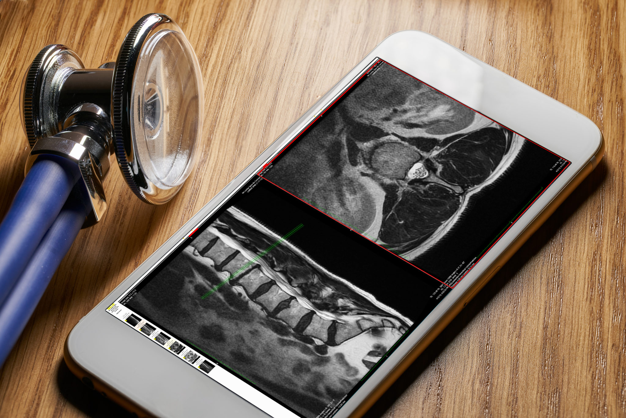
Without the science of radiology, doctors & specialists would not have as much detailed knowledge as to what is going on in the body. going on in the body.
X-rays, ultrasounds, scans and more are able to provide detailed images that take reveal what is behind a bad back, persistent headaches, or even cancer pain.
Crucial information allows better treatment and, ultimately, better healthcare. It gives the doctor and the patient the power of knowledge.
Radiology Services in Byron Bay
The North Coast Radiology Group is a longstanding member of the Northern Rivers healthcare community, recognised for excellence in providing consistent, caring and professional services to referring practitioners and patients utilising advanced, state of the art imaging technology.
All medical imaging modalities are represented by the Group including multi-slice CT, ultrasound, licensed MRI, nuclear medicine, general and dental radiography, mammography and bone densitometry. A large interventional service is also offered.
Radiology Services available at First Light Healthcare
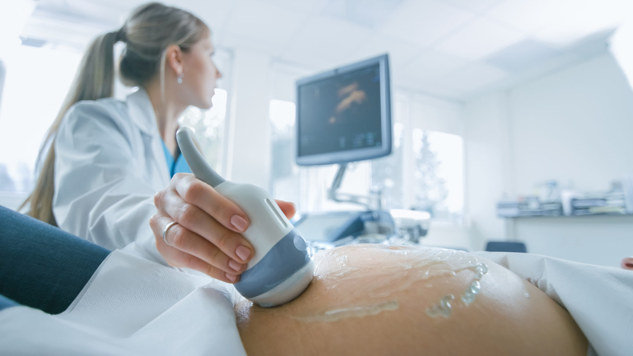
Ultrasound
For an ultrasound scan, high frequency sound waves are used to generate detailed images of the body.
First water-based gel is applied to your skin, then a highly trained Sonographer moves a hand-held ultrasound device over the area of interest.
Because no ionising radiation is used, ultrasounds are considered very safe, especially for pregnant women.
After the procedure, images are forwarded to specialist doctors to interpret while the technician prepares a report for the referring practitioner.
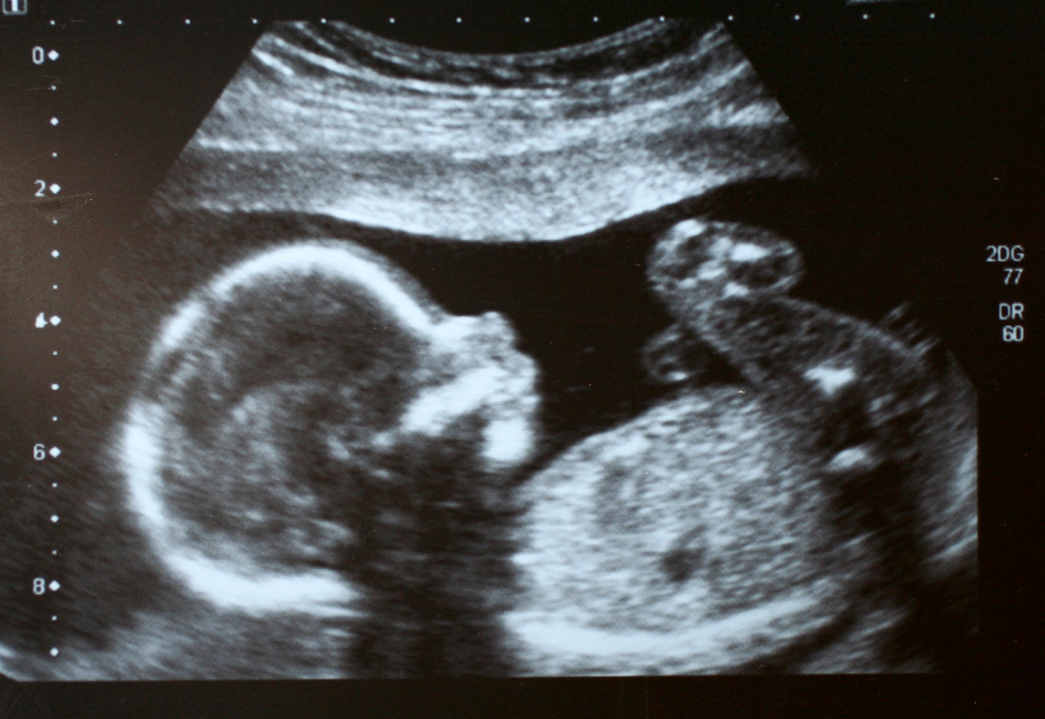
Pregnancy Scans
In the early stages of pregnancy (less than 12 weeks) an ultrasound may be performed if there has been bleeding or pain, if there is uncertainly about the date of the last period, or if there is excessive nausea or vomiting.
The Sonographer is able to measure the length of the fetus to estimate the stage of pregnancy and the due date of delivery. The heartbeat can be seen at about six weeks.
Around 20 weeks into pregnancy, a doctor will usually send an expectant mother for an ultrasound to check all is well.
The Sonographer checks that the placenta is developing normally and takes measurements of the fetus.
Patients receive a CD with these images to keep, free of charge.
They may need another ultrasound later if there has been difficulty in seeing the fetus or if the GP or obstetrician wants to check all is well.
Vascular Ultrasound
A vascular ultrasound, also called a duplex or Doppler scan, is an ultrasound that examines the blood vessels of the body.
Like all ultrasounds, it is a safe and non-invasive scan that is able to provide detailed, real time information without the use of radiation.
Nuchal Translucency
The Nuchal Translucency is the name for the normal fluid space behind the neck of the fetus that can be seen on ultrasound scans between 11 and 13 weeks.
An increase in the amount of fluid present may occur in the presence of certain chromosomal or structural abnormalities.
The Nuchal Translucency scan is part of the Combined First Trimester Screening Test, used to determine the risk of certain chromosomal abnormalities including Down Syndrome, and combines the patient’s medical history, an ultrasound and blood tests.
During the scan the Sonographer also looks at fetal size, heart rate and other anatomical structures.
The ultrasound data is entered into the Foetal Medicine Foundation database to calculate risk of certain abnormalities. If a risk is identified, this does not necessarily mean an abnormality is present. However, further testing will be required.
Questions and Answers
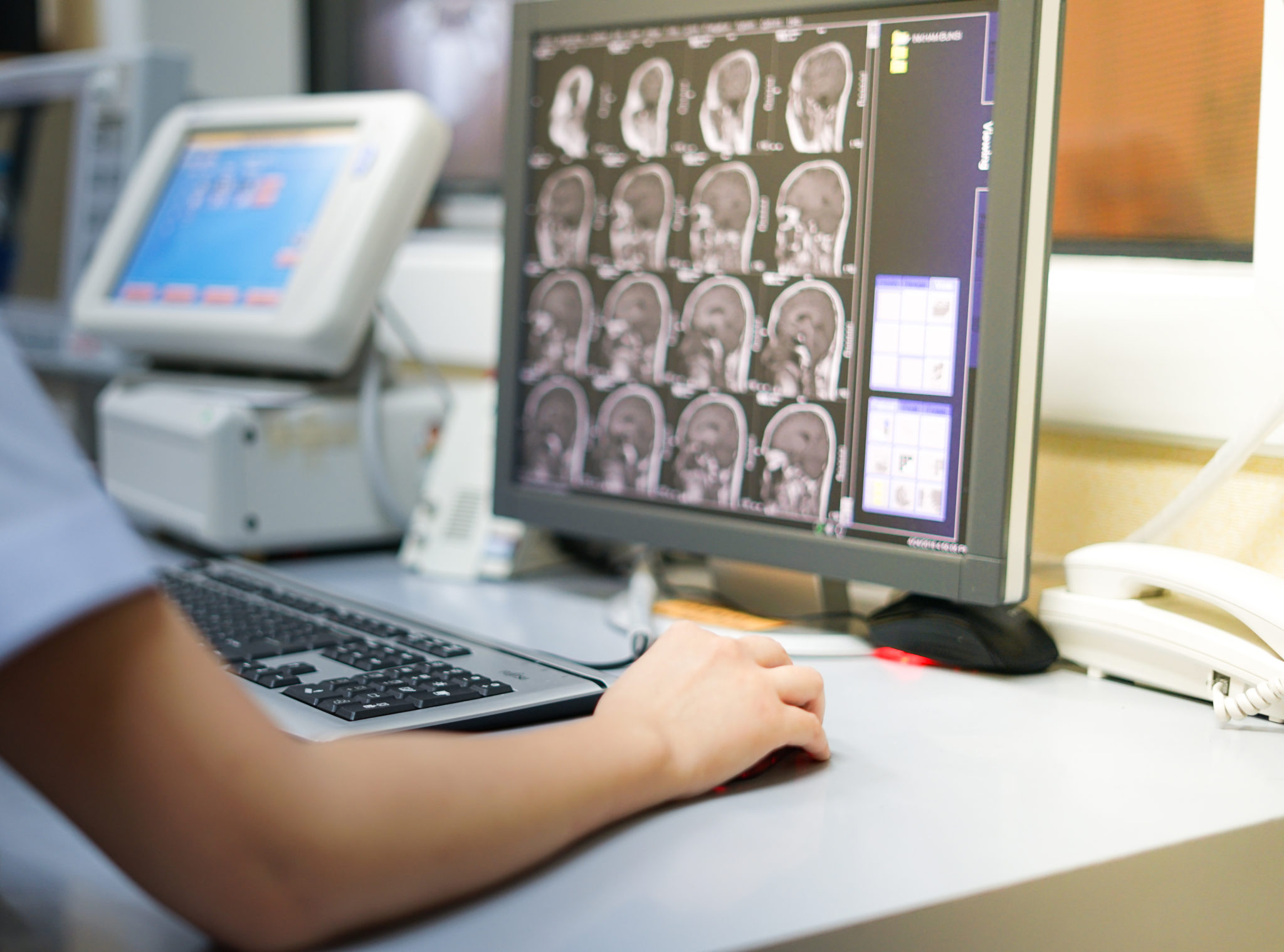
What is radiology?
Radiology is a specialist area of medicine involving tests such as X-rays, ultrasounds and scans.
Through the images that result, it is possible to see what is going on in the body, with organs, blood vessels, joints and more.
You will be referred for a radiology test, including an X-ray, scan or ultrasound, by your practitioner so that he or she may get a greater understanding of how to help you manage your health.

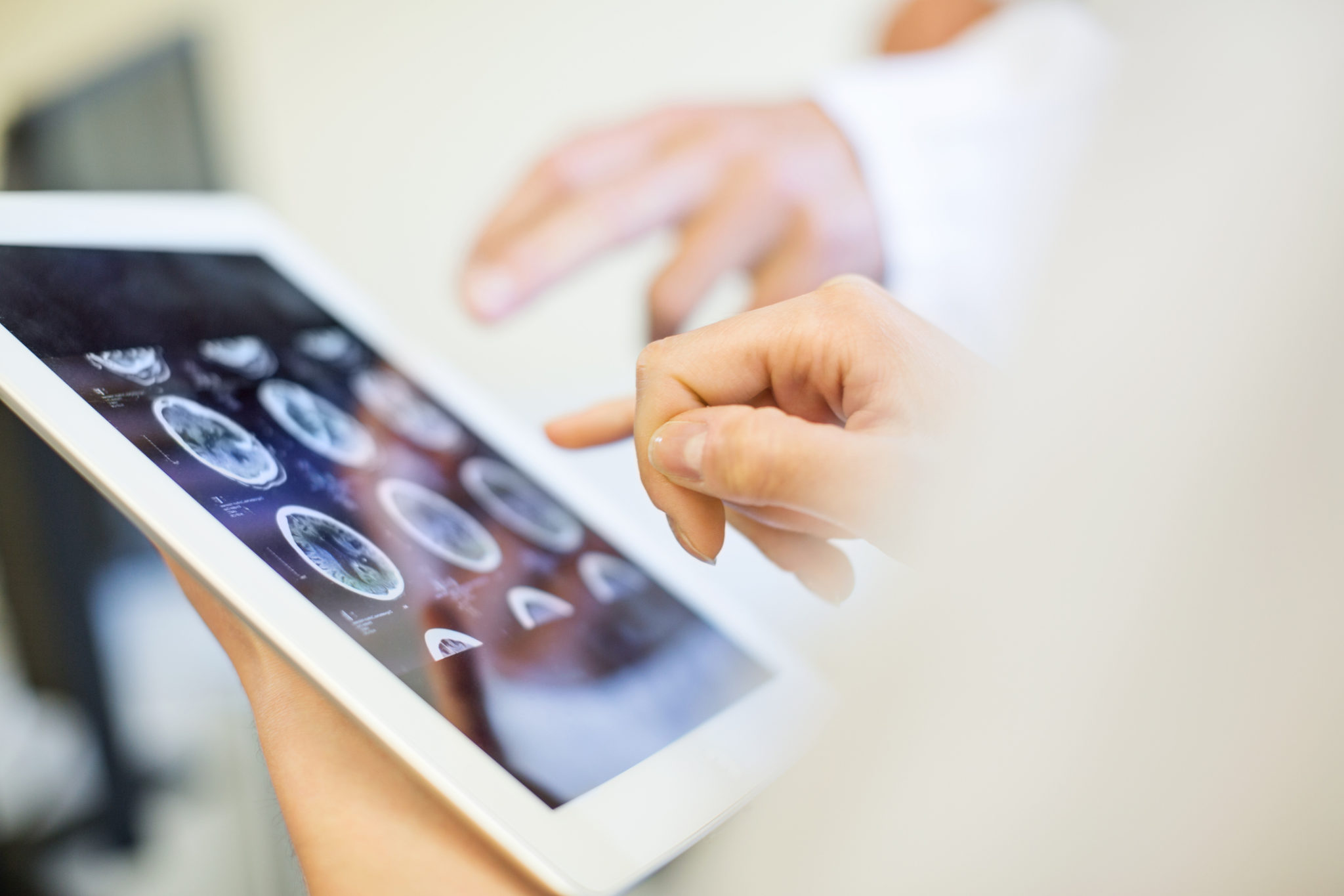
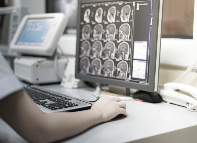
What is a radiologist? What does a clinical radiologist do?
A radiologist is a specialist medical doctor who has had postgraduate training in performing and interpreting diagnostic imaging tests.
He or she is fully trained in carrying our scans including X-ray, ultrasound, Computed Tomography scans and magnetic resonance imaging.
Using visual clues, a radiology is able to help your GP or specialist to understand symptoms, abnormalities or disease inside the body.
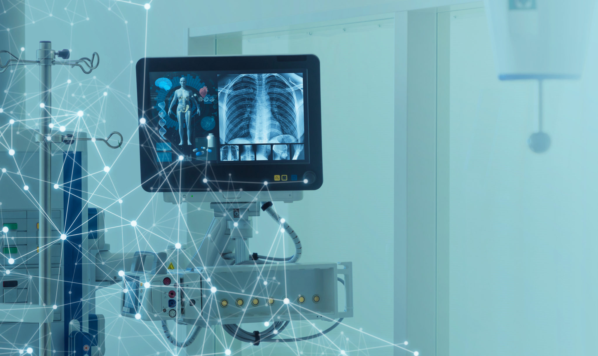
What can radiology detect?
Radiology can detect everything from a broken bone to a joint abnormality to a blood clot.
It can also be used to see how your body is healing, or responding to a treatment.
Diagnostic radiology is also used to screen for diseases, for example, for breast cancer and other cancers, or heart disease.
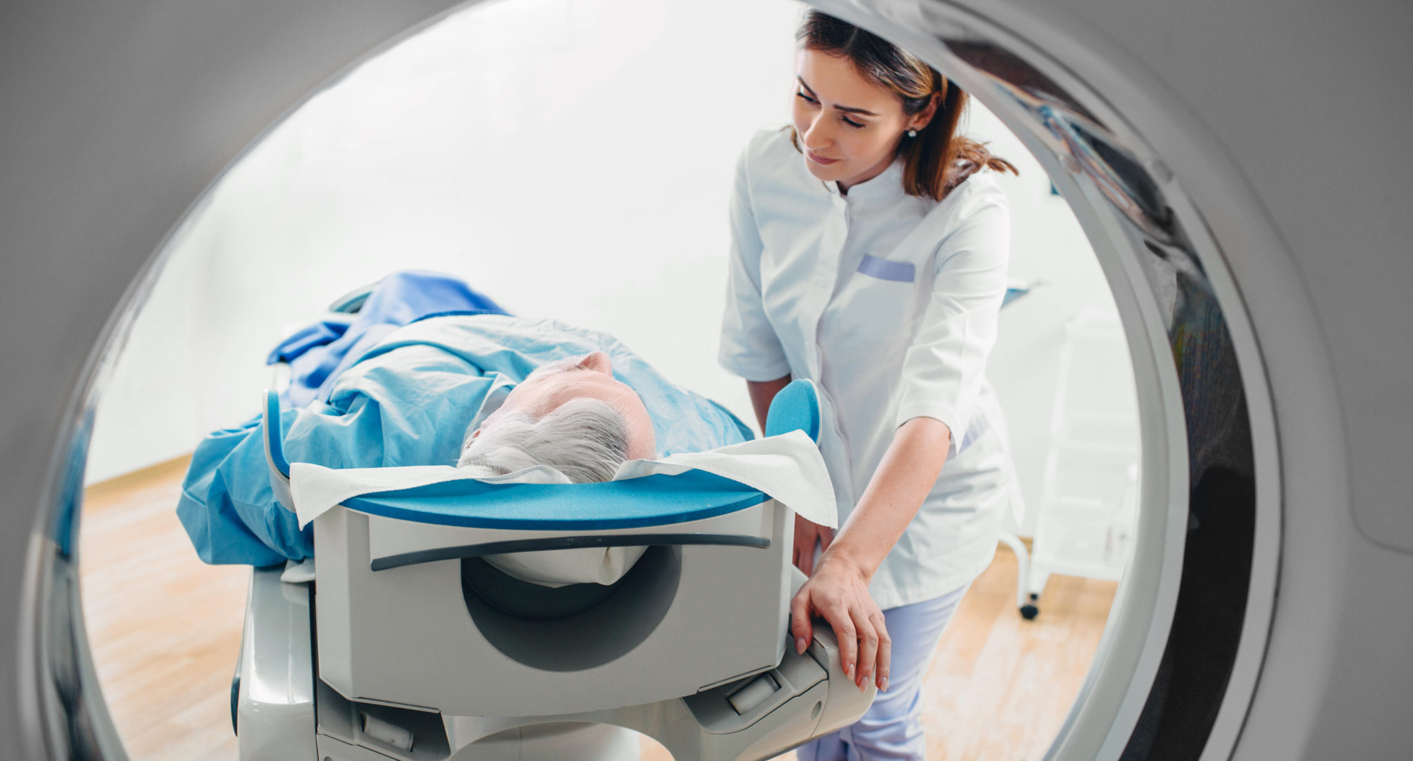
What are radiology tests?
Most people have heard of X-rays and, most have had one, even if that’s at the dentist.
But diagnostic radiology can also include ultrasound, CT scans, MRIs, and Nuclear Medicine Scans.
Some of these tests, for example a CT scan, an X-Ray and a mammogram, use a low dose or radiation to
What’s the difference between Radiology and Radiography?
Radiogaphy refers to the actual imaging tests. Radiology includes both the tests and treatments, such as radiation therapy.